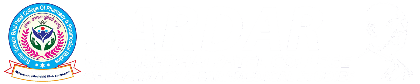
B.Voc in Radiology & Medical Imaging is a three-year undergraduate skill-based program designed to prepare students for careers in diagnostic imaging. The course focuses on training students to operate imaging machines such as X-ray, CT scan, MRI, and ultrasound to help diagnose diseases and monitor treatments. With the rise in demand for non-invasive diagnostic techniques, radiology professionals are needed in every modern hospital and diagnostic center. This program combines theory and practical knowledge, enabling students to work with doctors, radiologists, and healthcare teams. It also emphasizes patient safety, radiation protection, image analysis, and the ethical handling of diagnostic procedures.
Course Details – B.Voc in Radiology & Medical Imaging
This 3-year program is divided into six semesters and includes both academic and hands-on clinical training. Students learn about different imaging modalities, including conventional radiography, digital imaging, CT, MRI, ultrasound, and fluoroscopy. The curriculum also covers human anatomy, physiology, radiographic physics, and patient care techniques. As students progress, they are trained in positioning patients, understanding imaging protocols, and interpreting radiographs under professional guidance. Real-time exposure through internships in diagnostic centers and hospitals helps them gain confidence and build real-world skills. By the end of the course, students are job-ready and eligible to work as radiology technicians, imaging assistants, or pursue further specialization.
Examination Pattern –B.Voc in Radiology & Medical Imaging
The examination structure of this program consists of theory exams, practical tests, and internal assessments. Each semester includes written papers on anatomy, radiographic physics, image processing, and patient care. Practical exams require students to demonstrate correct usage of machines like X-ray and simulate clinical procedures. Viva voce and assignments test their understanding of safety norms, imaging techniques, and emergency handling. Internal marks are awarded based on attendance, practical records, behavior, and performance during clinical postings. The balanced exam pattern ensures students excel in both theoretical knowledge and applied diagnostic skills essential for real-world healthcare environments.
Syllabus Introduction
The syllabus is carefully designed to provide deep understanding of medical imaging sciences along with radiation safety, patient care, and image evaluation. The curriculum builds strong technical foundations while preparing students for advanced imaging technologies used in modern hospitals.
Year 1: Basics of Imaging & Anatomy
Human Anatomy & Physiology
Introduction to Radiology
Radiographic Physics – I
Radiation Hazards & Safety
Patient Care & Communication
Environmental Science
Basic Computer Applications
Year 2: Core Radiographic Techniques
Radiographic Physics – II
General Radiography Procedures
Radiographic Positioning Techniques
Image Processing & Darkroom Techniques
Medical Ethics and Legal Aspects
Clinical Posting – I
Pathology Basics
Year 3: Advanced Imaging & Clinical Training
Advanced Imaging Techniques (CT, MRI, Ultrasound)
Radiographic Quality Control
Hospital Internship / Diagnostic Center Posting
Equipment Maintenance and Troubleshooting
Emergency Radiology Procedures
Project Work & Case Study
Additional Highlights:
Hands-on practice in X-ray, CT, and MRI machines
Internship opportunities in government and private diagnostic centers
Focus on patient positioning, radiation safety, and image clarity
Exposure to PACS (Picture Archiving and Communication Systems)